General Information About Melanoma
Melanoma is a disease in which malignant (cancer) cells form in melanocytes (cells that color the skin).
The skin is the body's largest organ. It protects against heat, sunlight, injury, and infection. Skin also helps control body temperature and stores water, fat, and vitamin D. The skin has several layers, but the two main layers are the epidermis (upper or outer layer) and the dermis (lower or inner layer). Skin cancer begins in the epidermis, which is made up of three kinds of cells:
- Squamous cells: Thin, flat cells that form the top layer of the epidermis.
- Basal cells: Round cells under the squamous cells.
- Melanocytes: Cells that make melanin and are found in the lower part of the epidermis. Melanin is the pigment that gives skin its natural color. When skin is exposed to the sun or artificial light, melanocytes make more pigment and cause the skin to darken.
The number of new cases of melanoma has been increasing over the last 40 years. Melanoma is most common in adults, but it is sometimes found in children and adolescents. (See the PDQ summary on Unusual Cancers of Childhood Treatment for more information on melanoma in children and adolescents.)
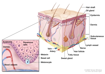
Anatomy of the skin, showing the epidermis, dermis, and subcutaneous tissue. Melanocytes are in the layer of basal cells at the deepest part of the epidermis.
There are different types of cancer that start in the skin.
There are two forms of skin cancer: melanoma and nonmelanoma.
Melanoma is a rare form of skin cancer. It is more likely to invade nearby tissues and spread to other parts of the body than other types of skin cancer. When melanoma starts in the skin, it is called cutaneous melanoma. Melanoma may also occur in mucous membranes (thin, moist layers of tissue that cover surfaces such as the lips). This PDQ summary is about cutaneous (skin) melanoma and melanoma that affects the mucous membranes.
The most common types of skin cancer are basal cell carcinoma and squamous cell carcinoma. They are nonmelanoma skin cancers. Nonmelanoma skin cancers rarely spread to other parts of the body. (See the PDQ summary on Skin Cancer Treatment for more information on basal cell and squamous cell skin cancer.)
Melanoma can occur anywhere on the skin.
In men, melanoma is often found on the trunk (the area from the shoulders to the hips) or the head and neck. In women, melanoma forms most often on the arms and legs.
When melanoma occurs in the eye, it is called intraocular or ocular melanoma. (See the PDQ summary on Intraocular (Uveal) Melanoma Treatment for more information.)
Unusual moles, exposure to sunlight, and health history can affect the risk of melanoma.
Anything that increases your risk of getting a disease is called a risk factor. Having a risk factor does not mean that you will get cancer; not having risk factors doesn't mean that you will not get cancer. Talk with your doctor if you think you may be at risk.
Risk factors for melanoma include the following:
- Having a fair complexion, which includes the following:
- Fair skin that freckles and burns easily, does not tan, or tans poorly.
- Blue or green or other light-colored eyes.
- Red or blond hair.
- Being exposed to natural sunlight or artificial sunlight (such as from tanning beds) over long periods of time.
- Being exposed to certain factors in the environment (in the air, your home or workplace, and your food and water). Some of the environmental risk factors for melanoma are radiation, solvents, vinyl chloride, and PCBs.
- Having a history of many blistering sunburns, especially as a child or teenager.
- Having several large or many small moles.
- Having a family history of unusual moles (atypical nevus syndrome).
- Having a family or personal history of melanoma.
- Being white.
- Having a weakened immune system.
- Having certain changes in the genes that are linked to melanoma.
Being white or having a fair complexion increases the risk of melanoma, but anyone can have melanoma, including people with dark skin.
See the following PDQ summaries for more information on risk factors for melanoma:
- Genetics of Skin Cancer
- Skin Cancer Prevention
Signs of melanoma include a change in the way a mole or pigmented area looks.
These and other signs and symptoms may be caused by melanoma or by other conditions. Check with your doctor if you have any of the following:
- A mole that:
- changes in size, shape, or color.
- has irregular edges or borders.
- is more than one color.
- is asymmetrical (if the mole is divided in half, the 2 halves are different in size or shape).
- itches.
- oozes, bleeds, or is ulcerated (a hole forms in the skin when the top layer of cells breaks down and the tissue below shows through).
- A change in pigmented (colored) skin.
- Satellite moles (new moles that grow near an existing mole).
For pictures and descriptions of common moles and melanoma, see Common Moles, Dysplastic Nevi, and Risk of Melanoma.
Tests that examine the skin are used to detect (find) and diagnose melanoma.
If a mole or pigmented area of the skin changes or looks abnormal, the following tests and procedures can help find and diagnose melanoma:
- Skin exam: A doctor or nurse checks the skin for moles, birthmarks, or other pigmented areas that look abnormal in color, size, shape, or texture.
- Biopsy: A procedure to remove the abnormal tissue and a small amount of normal tissue around it. A pathologist looks at the tissue under a microscope to check for cancer cells. It can be hard to tell the difference between a colored mole and an early melanoma lesion. Patients may want to have the sample of tissue checked by a second pathologist. If the abnormal mole or lesion is cancer, the sample of tissue may also be tested for certain gene changes.
It is important that abnormal areas of the skin not be shaved off or cauterized (destroyed with a hot instrument, an electric current, or a caustic substance) because cancer cells that remain may grow and spread.
See the PDQ summary on Skin Cancer Screening for more information.
Certain factors affect prognosis (chance of recovery) and treatment options.
The prognosis (chance of recovery) and treatment options depend on the following:
- The thickness of the tumor and where it is in the body.
- How quickly the cancer cells are dividing.
- Whether there was bleeding or ulceration of the tumor.
- How much cancer is in the lymph nodes.
- The number of places cancer has spread to in the body.
- The level of lactate dehydrogenase (LDH) in the blood.
- Whether the cancer has certain mutations (changes) in a gene called BRAF.
- The patient's age and general health.
Stages of Melanoma
After melanoma has been diagnosed, tests are done to find out if cancer cells have spread within the skin or to other parts of the body.
The process used to find out whether cancer has spread within the skin or to other parts of the body is called staging. The information gathered from the staging process determines the stage of the disease. It is important to know the stage in order to plan treatment.
The following tests and procedures may be used in the staging process:
- Physical exam and history: An exam of the body to check general signs of health, including checking for signs of disease, such as lumps or anything else that seems unusual. A history of the patient's health habits and past illnesses and treatments will also be taken.
- Lymph node mapping and sentinel lymph node biopsy: Procedures in which a radioactive substance and/or blue dye is injected near the tumor. The substance or dye flows through lymph ducts to the sentinel node or nodes (the first lymph node or nodes where cancer cells are likely to spread). The surgeon removes only the nodes with the radioactive substance or dye. A pathologist views a sample of tissue under a microscope to check for cancer cells. If no cancer cells are found, it may not be necessary to remove more nodes.
- CT scan (CAT scan): A procedure that makes a series of detailed pictures of areas inside the body taken from different angles. The pictures are made by a computer linked to an x-ray machine. A dye may be injected into a vein or swallowed to help the organs or tissues show up more clearly. This procedure is also called computed tomography, computerized tomography, or computerized axial tomography. For melanoma, pictures may be taken of the chest, abdomen, and pelvis.
- PET scan (positron emission tomography scan): A procedure to find malignant tumor cells in the body. A small amount of radioactive glucose (sugar) is injected into a vein. The PET scanner rotates around the body and makes a picture of where glucose is being used in the body. Malignant tumor cells show up brighter in the picture because they are more active and take up more glucose than normal cells do.
- MRI (magnetic resonance imaging) with gadolinium: A procedure that uses a magnet, radio waves, and a computer to make a series of detailed pictures of areas inside the body, such as the brain. A substance called gadolinium is injected into a vein. The gadolinium collects around the cancer cells so they show up brighter in the picture. This procedure is also called nuclear magnetic resonance imaging (NMRI).
- Blood chemistry studies: A procedure in which a blood sample is checked to measure the amounts of certain substances released into the blood by organs and tissues in the body. For melanoma, the blood is checked for an enzyme called lactate dehydrogenase (LDH). LDH levels that are higher than normal may be a sign of melanoma.
The results of these tests are viewed together with the results of the tumor biopsy to find out the stage of the melanoma.
There are three ways that cancer spreads in the body.
Cancer can spread through tissue, the lymph system, and the blood:
- Tissue. The cancer spreads from where it began by growing into nearby areas.
- Lymph system. The cancer spreads from where it began by getting into the lymph system. The cancer travels through the lymph vessels to other parts of the body.
- Blood. The cancer spreads from where it began by getting into the blood. The cancer travels through the blood vessels to other parts of the body.
Cancer may spread from where it began to other parts of the body.
When cancer spreads to another part of the body, it is called metastasis. Cancer cells break away from where they began (the primary tumor) and travel through the lymph system or blood.
- Lymph system. The cancer gets into the lymph system, travels through the lymph vessels, and forms a tumor (metastatic tumor) in another part of the body.
- Blood. The cancer gets into the blood, travels through the blood vessels, and forms a tumor (metastatic tumor) in another part of the body.
The metastatic tumor is the same type of cancer as the primary tumor. For example, if melanoma spreads to the lung, the cancer cells in the lung are actually melanoma cells. The disease is metastatic melanoma, not lung cancer.
The method used to stage melanoma is based mainly on the thickness of the tumor and whether cancer has spread to lymph nodes or other parts of the body.
The staging of melanoma depends on the following:
- The thickness of the tumor. The thickness is described using the Breslow scale.
- Whether the tumor is ulcerated (has broken through the skin).
- Whether the tumor has spread to the lymph nodes and if the lymph nodes are joined together (matted).
- Whether the tumor has spread to other parts of the body.
The following stages are used for melanoma:
Stage 0 (Melanoma in Situ)
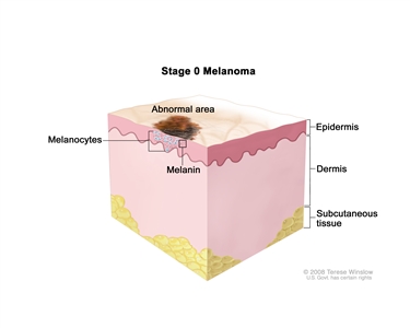
Stage 0 melanoma in situ. Abnormal melanocytes are in the epidermis (outer layer of the skin).
In stage 0, abnormal melanocytes are found in the epidermis. These abnormal melanocytes may become cancer and spread into nearby normal tissue. Stage 0 is also called melanoma in situ.
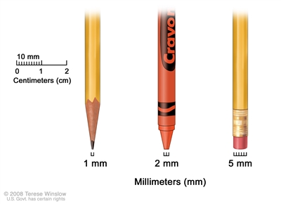
Millimeters (mm). A sharp pencil point is about 1 mm, a new crayon point is about 2 mm, and a new pencil eraser is about 5 mm.
Stage I
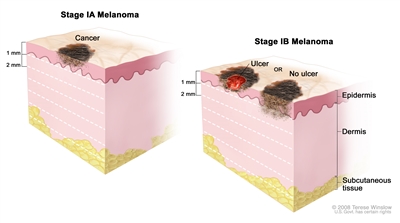
Stage I melanoma. In stage IA, the tumor is not more than 1 millimeter thick, with no ulceration (break in the skin). In stage IB, the tumor is either not more than 1 millimeter thick, with ulceration, OR more than 1 but not more than 2 millimeters thick, with no ulceration. Skin thickness is different on different parts of the body.
In stage I, cancer has formed. Stage I is divided into stages IA and IB.
- Stage IA: In stage IA, the tumor is not more than 1 millimeter thick, with no ulceration.
- Stage IB: In stage IB, the tumor is either:
- not more than 1 millimeter thick and it has ulceration; or
- more than 1 but not more than 2 millimeters thick, with no ulceration.
Stage II
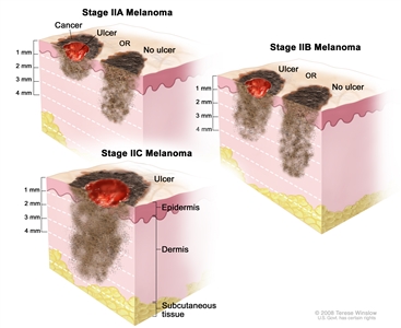
Stage II melanoma. In stage IIA, the tumor is either more than 1 but not more than 2 millimeters thick, with ulceration (break in the skin), OR it is more than 2 but not more than 4 millimeters thick, with no ulceration. In stage IIB, the tumor is either more than 2 but not more than 4 millimeters thick, with ulceration, OR it is more than 4 millimeters thick, with no ulceration. In stage IIC, the tumor is more than 4 millimeters thick, with ulceration. Skin thickness is different on different parts of the body.
Stage II is divided into stages IIA, IIB, and IIC.
- Stage IIA: In stage IIA, the tumor is either:
- more than 1 but not more than 2 millimeters thick, with ulceration; or
- more than 2 but not more than 4 millimeters thick, with no ulceration.
- Stage IIB: In stage IIB, the tumor is either:
- more than 2 but not more than 4 millimeters thick, with ulceration; or
- more than 4 millimeters thick, with no ulceration.
- Stage IIC: In stage IIC, the tumor is more than 4 millimeters thick, with ulceration.
Stage III
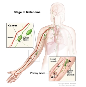
Stage III melanoma. The tumor may be any thickness, with or without ulceration (a break in the skin), and (a) cancer has spread to one or more lymph nodes; (b) lymph nodes with cancer may be joined together (matted); (c) cancer may be in a lymph vessel between the primary tumor and nearby lymph nodes; and/or (d) very small tumors may be found on or under the skin, not more than 2 centimeters away from the primary tumor.
In stage III, the tumor may be any thickness, with or without ulceration. One or more of the following is true:
- Cancer has spread to one or more lymph nodes.
- Lymph nodes are joined together (matted).
- Cancer is in a lymph vessel between the primary tumor and nearby lymph nodes. The cancer is more than 2 centimeters away from the primary tumor.
- Very small tumors are found on or under the skin, not more than 2 centimeters away from the primary tumor.
Stage IV
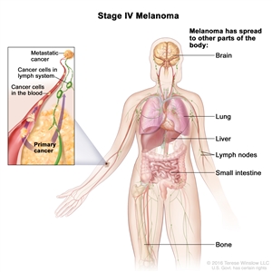
Stage IV melanoma. The cancer has spread to other parts of the body, such as the brain, lung, liver, lymph nodes, small intestine, and bone.
In stage IV, the cancer has spread to other places in the body, such as the lung, liver, brain, bone, soft tissue, or gastrointestinal (GI) tract. Cancer may have spread to places in the skin far away from where it first started.
Recurrent Melanoma
Recurrent melanoma is cancer that has recurred (come back) after it has been treated. The cancer may come back in the area where it first started or in other parts of the body, such as the lungs or liver.
Treatment Option Overview
There are different types of treatment for patients with melanoma.
Different types of treatment are available for patients with melanoma. Some treatments are standard (the currently used treatment), and some are being tested in clinical trials. A treatment clinical trial is a research study meant to help improve current treatments or obtain information on new treatments for patients with cancer. When clinical trials show that a new treatment is better than the standard treatment, the new treatment may become the standard treatment. Patients may want to think about taking part in a clinical trial. Some clinical trials are open only to patients who have not started treatment.
Five types of standard treatment are used:
Surgery
Surgery to remove the tumor is the primary treatment of all stages of melanoma. A wide local excision is used to remove the melanoma and some of the normal tissue around it. Skin grafting (taking skin from another part of the body to replace the skin that is removed) may be done to cover the wound caused by surgery.
It is important to know whether cancer has spread to the lymph nodes. Lymph node mapping and sentinel lymph node biopsy are done to check for cancer in the sentinel lymph node (the first lymph node the cancer is likely to spread to from the tumor) during surgery. A radioactive substance and/or blue dye is injected near the tumor. The substance or dye flows through the lymph ducts to the lymph nodes. The first lymph node to receive the substance or dye is removed. A pathologist views the tissue under a microscope to look for cancer cells. If cancer cells are found, more lymph nodes will be removed and tissue samples will be checked for signs of cancer. This is called a lymphadenectomy.
Even if the doctor removes all the melanoma that can be seen at the time of surgery, some patients may be given chemotherapy after surgery to kill any cancer cells that are left. Chemotherapy given after surgery, to lower the risk that the cancer will come back, is called adjuvant therapy.
Surgery to remove cancer that has spread to the lymph nodes, lung, gastrointestinal (GI) tract, bone, or brain may be done to improve the patient's quality of life by controlling symptoms.
Chemotherapy
Chemotherapy is a cancer treatment that uses drugs to stop the growth of cancer cells, either by killing the cells or by stopping them from dividing. When chemotherapy is taken by mouth or injected into a vein or muscle, the drugs enter the bloodstream and can reach cancer cells throughout the body (systemic chemotherapy). When chemotherapy is placed directly into the cerebrospinal fluid, an organ, or a body cavity such as the abdomen, the drugs mainly affect cancer cells in those areas (regional chemotherapy).
One type of regional chemotherapy is hyperthermic isolated limb perfusion. With this method, anticancer drugs go directly to the arm or leg the cancer is in. The flow of blood to and from the limb is temporarily stopped with a tourniquet. A warm solution with the anticancer drug is put directly into the blood of the limb. This gives a high dose of drugs to the area where the cancer is.
The way the chemotherapy is given depends on the type and stage of the cancer being treated.
See Drugs Approved for Melanoma for more information.
Radiation therapy
Radiation therapy is a cancer treatment that uses high-energy x-rays or other types of radiation to kill cancer cells or keep them from growing. There are two types of radiation therapy:
- External radiation therapy uses a machine outside the body to send radiation toward the cancer.
- Internal radiation therapy uses a radioactive substance sealed in needles, seeds, wires, or catheters that are placed directly into or near the cancer.
The way the radiation therapy is given depends on the type and stage of the cancer being treated. External radiation therapy is used to treat melanoma, and may also be used as palliative therapy to relieve symptoms and improve quality of life.
Immunotherapy
Immunotherapy is a treatment that uses the patient's immune system to fight cancer. Substances made by the body or made in a laboratory are used to boost, direct, or restore the body's natural defenses against cancer. This type of cancer treatment is also called biotherapy or biologic therapy.
The following types of immunotherapy are being used in the treatment of melanoma:
See Drugs Approved for Melanoma for more information.
Targeted therapy
Targeted therapy is a type of treatment that uses drugs or other substances to attack cancer cells. Targeted therapies usually cause less harm to normal cells than chemotherapy or radiation therapy do. The following types of targeted therapy are used or being studied in the treatment of melanoma:
- Signal transduction inhibitor therapy: Signal transduction inhibitors block signals that are passed from one molecule to another inside a cell. Blocking these signals may kill cancer cells.
- Vemurafenib, dabrafenib, trametinib, and cobimetinib are signal transduction inhibitors used to treat some patients with advanced melanoma or tumors that cannot be removed by surgery. Vemurafenib and dabrafenib block the activity of proteins made by mutant BRAF genes. Trametinib and cobimetinib affect the growth and survival of cancer cells.
- Oncolytic virus therapy: A type of targeted therapy that is used in the treatment of melanoma. Oncolytic virus therapy uses a virus that infects and breaks down cancer cells but not normal cells. Radiation therapy or chemotherapy may be given after oncolytic virus therapy to kill more cancer cells.
- Angiogenesis inhibitors: A type of targeted therapy that is being studied in the treatment of melanoma. Angiogenesis inhibitors block the growth of new blood vessels. In cancer treatment, they may be given to prevent the growth of new blood vessels that tumors need to grow.
New targeted therapies and combinations of therapies are being studied in the treatment of melanoma.
See Drugs Approved for Melanoma for more information.
New types of treatment are being tested in clinical trials.
Information about clinical trials is available from the NCI website
Patients may want to think about taking part in a clinical trial.
For some patients, taking part in a clinical trial may be the best treatment choice. Clinical trials are part of the cancer research process. Clinical trials are done to find out if new cancer treatments are safe and effective or better than the standard treatment.
Many of today's standard treatments for cancer are based on earlier clinical trials. Patients who take part in a clinical trial may receive the standard treatment or be among the first to receive a new treatment.
Patients who take part in clinical trials also help improve the way cancer will be treated in the future. Even when clinical trials do not lead to effective new treatments, they often answer important questions and help move research forward.
Patients can enter clinical trials before, during, or after starting their cancer treatment.
Some clinical trials only include patients who have not yet received treatment. Other trials test treatments for patients whose cancer has not gotten better. There are also clinical trials that test new ways to stop cancer from recurring (coming back) or reduce the side effects of cancer treatment.
Clinical trials are taking place in many parts of the country. See the Treatment Options section that follows for links to current treatment clinical trials. These have been retrieved from NCI's listing of clinical trials.
Follow-up tests may be needed.
Some of the tests that were done to diagnose the cancer or to find out the stage of the cancer may be repeated. Some tests will be repeated in order to see how well the treatment is working. Decisions about whether to continue, change, or stop treatment may be based on the results of these tests.
Some of the tests will continue to be done from time to time after treatment has ended. The results of these tests can show if your condition has changed or if the cancer has recurred (come back). These tests are sometimes called follow-up tests or check-ups.
Treatment Options by Stage
Stage 0 (Melanoma in Situ)
Treatment of stage 0 is usually surgery to remove the area of abnormal cells and a small amount of normal tissue around it.
Check the list of NCI-supported cancer clinical trials that are now accepting patients with stage 0 melanoma. For more specific results, refine the search by using other search features, such as the location of the trial, the type of treatment, or the name of the drug. Talk with your doctor about clinical trials that may be right for you. General information about clinical trials is available from the NCI website.
Stage I Melanoma
Treatment of stage I melanoma may include the following:
- Surgery to remove the tumor and some of the normal tissue around it. Sometimes lymph node mapping and removal of lymph nodes is also done.
- A clinical trial of new ways to find cancer cells in the lymph nodes.
Check the list of NCI-supported cancer clinical trials that are now accepting patients with stage I melanoma. For more specific results, refine the search by using other search features, such as the location of the trial, the type of treatment, or the name of the drug. Talk with your doctor about clinical trials that may be right for you. General information about clinical trials is available from the NCI website.
Stage II Melanoma
Treatment of stage II melanoma may include the following:
- Surgery to remove the tumor and some of the normal tissue around it. Sometimes lymph node mapping and sentinel lymph node biopsy are done to check for cancer in the lymph nodes at the same time as the surgery to remove the tumor. If cancer is found in the sentinel lymph node, more lymph nodes may be removed.
- Surgery followed by immunotherapy with interferon if there is a high risk that the cancer will come back.
- A clinical trial of new types of treatment to be used after surgery.
Check the list of NCI-supported cancer clinical trials that are now accepting patients with stage II melanoma. For more specific results, refine the search by using other search features, such as the location of the trial, the type of treatment, or the name of the drug. Talk with your doctor about clinical trials that may be right for you. General information about clinical trials is available from the NCI website.
Stage III Melanoma That Can Be Removed By Surgery
Treatment of stage III melanoma that can be removed by surgery may include the following:
- Surgery to remove the tumor and some of the normal tissue around it. Skin grafting may be done to cover the wound caused by surgery. Sometimes lymph node mapping and sentinel lymph node biopsy are done to check for cancer in the lymph nodes at the same time as the surgery to remove the tumor. If cancer is found in the sentinel lymph node, more lymph nodes may be removed.
- Surgery followed by immunotherapy with ipilimumab or interferon if there is a high risk that the cancer will come back.
- A clinical trial of immunotherapy or targeted therapy to be used after surgery.
Check the list of NCI-supported cancer clinical trials that are now accepting patients with stage III melanoma. For more specific results, refine the search by using other search features, such as the location of the trial, the type of treatment, or the name of the drug. Talk with your doctor about clinical trials that may be right for you. General information about clinical trials is available from the NCI website.
Stage III Melanoma That Cannot Be Removed By Surgery, Stage IV Melanoma, and Recurrent Melanoma
Treatment of stage III melanoma that cannot be removed by surgery, stage IV melanoma, and recurrent melanoma may include the following:
- Immunotherapy with ipilimumab, pembrolizumab, nivolumab, or interleukin-2 (IL-2). Sometimes ipilimumab and nivolumab are given together.
- Targeted therapy with vemurafenib, dabrafenib, trametinib, or cobimetinib. Sometimes vemurafenib and cobimetinib or dabrafenib and trametinib are given together.
- Injections into the tumor, such as oncolytic virus therapy.
- Chemotherapy.
- Palliative therapy to relieve symptoms and improve the quality of life. This may include:
- Surgery to remove lymph nodes or tumors in the lung, gastrointestinal (GI) tract, bone, or brain.
- Radiation therapy to the brain, spinal cord, or bone.
Treatments that are being studied in clinical trials for stage III melanoma that cannot be removed by surgery, stage IV melanoma, and recurrent melanoma include the following:
- Immunotherapy alone or in combination with other therapies such as targeted therapy.
- Targeted therapy, such as signal transduction inhibitors, angiogenesis inhibitors, oncolytic virus therapy, or drugs that target certain gene mutations. These may be given alone or in combination.
- Surgery to remove all known cancer.
- Regional chemotherapy (hyperthermic isolated limb perfusion). Some patients may also have immunotherapy with tumor necrosis factor.
- Systemic chemotherapy.
Check the list of NCI-supported cancer clinical trials that are now accepting patients with stage IV melanoma and recurrent melanoma. For more specific results, refine the search by using other search features, such as the location of the trial, the type of treatment, or the name of the drug. Talk with your doctor about clinical trials that may be right for you. General information about clinical trials is available from the NCI website.
To Learn More About Melanoma
For more information from the National Cancer Institute about melanoma, see the following:
- Skin Cancer (Including Melanoma) Home Page
- Skin Cancer Prevention
- Skin Cancer Screening
- Sentinel Lymph Node Biopsy
- Drugs Approved for Melanoma
- Biological Therapies for Cancer
- Targeted Cancer Therapies
- Moles to Melanoma: Recognizing the ABCDE Features
For general cancer information and other resources from the National Cancer Institute, see the following:
- About Cancer
- Staging
- Chemotherapy and You: Support for People With Cancer
- Radiation Therapy and You: Support for People With Cancer
- Coping with Cancer
- Questions to Ask Your Doctor about Cancer
- For Survivors and Caregivers
About This PDQ Summary
About PDQ
Physician Data Query (PDQ) is the National Cancer Institute's (NCI's) comprehensive cancer information database. The PDQ database contains summaries of the latest published information on cancer prevention, detection, genetics, treatment, supportive care, and complementary and alternative medicine. Most summaries come in two versions. The health professional versions have detailed information written in technical language. The patient versions are written in easy-to-understand, nontechnical language. Both versions have cancer information that is accurate and up to date and most versions are also available in Spanish.
PDQ is a service of the NCI. The NCI is part of the National Institutes of Health (NIH). NIH is the federal government's center of biomedical research. The PDQ summaries are based on an independent review of the medical literature. They are not policy statements of the NCI or the NIH.
Purpose of This Summary
This PDQ cancer information summary has current information about the treatment of melanoma. It is meant to inform and help patients, families, and caregivers. It does not give formal guidelines or recommendations for making decisions about health care.
Reviewers and Updates
Editorial Boards write the PDQ cancer information summaries and keep them up to date. These Boards are made up of experts in cancer treatment and other specialties related to cancer. The summaries are reviewed regularly and changes are made when there is new information. The date on each summary ("Date Last Modified") is the date of the most recent change.
The information in this patient summary was taken from the health professional version, which is reviewed regularly and updated as needed, by the PDQ Adult Treatment Editorial Board.
Clinical Trial Information
A clinical trial is a study to answer a scientific question, such as whether one treatment is better than another. Trials are based on past studies and what has been learned in the laboratory. Each trial answers certain scientific questions in order to find new and better ways to help cancer patients. During treatment clinical trials, information is collected about the effects of a new treatment and how well it works. If a clinical trial shows that a new treatment is better than one currently being used, the new treatment may become "standard." Patients may want to think about taking part in a clinical trial. Some clinical trials are open only to patients who have not started treatment.
Clinical trials are listed in PDQ and can be found online at NCI's website. Many cancer doctors who take part in clinical trials are also listed in PDQ. For more information, call the Cancer Information Service 1-800-4-CANCER (1-800-422-6237).
Permission to Use This Summary
PDQ is a registered trademark. The content of PDQ documents can be used freely as text. It cannot be identified as an NCI PDQ cancer information summary unless the whole summary is shown and it is updated regularly. However, a user would be allowed to write a sentence such as "NCI's PDQ cancer information summary about breast cancer prevention states the risks in the following way: [include excerpt from the summary]."
The best way to cite this PDQ summary is:
PDQ® Adult Treatment Editorial Board. PDQ Melanoma Treatment. Bethesda, MD: National Cancer Institute. Updated <MM/DD/YYYY>. Available at: https://www.cancer.gov/types/skin/patient/melanoma-treatment-pdq. Accessed <MM/DD/YYYY>. [PMID: 26389388]
Images in this summary are used with permission of the author(s), artist, and/or publisher for use in the PDQ summaries only. If you want to use an image from a PDQ summary and you are not using the whole summary, you must get permission from the owner. It cannot be given by the National Cancer Institute. Information about using the images in this summary, along with many other images related to cancer can be found in Visuals Online. Visuals Online is a collection of more than 2,000 scientific images.
Disclaimer
The information in these summaries should not be used to make decisions about insurance reimbursement. More information on insurance coverage is available on Cancer.gov on the Managing Cancer Care page.
Contact Us
More information about contacting us or receiving help with the Cancer.gov website can be found on our Contact Us for Help page. Questions can also be submitted to Cancer.gov through the website's E-mail Us.
Last Revised: 2017-03-10
If you want to know more about cancer and how it is treated, or if you wish to know about clinical trials for your type of cancer, you can call the NCI's Cancer Information Service at 1-800-422-6237, toll free. A trained information specialist can talk with you and answer your questions.